
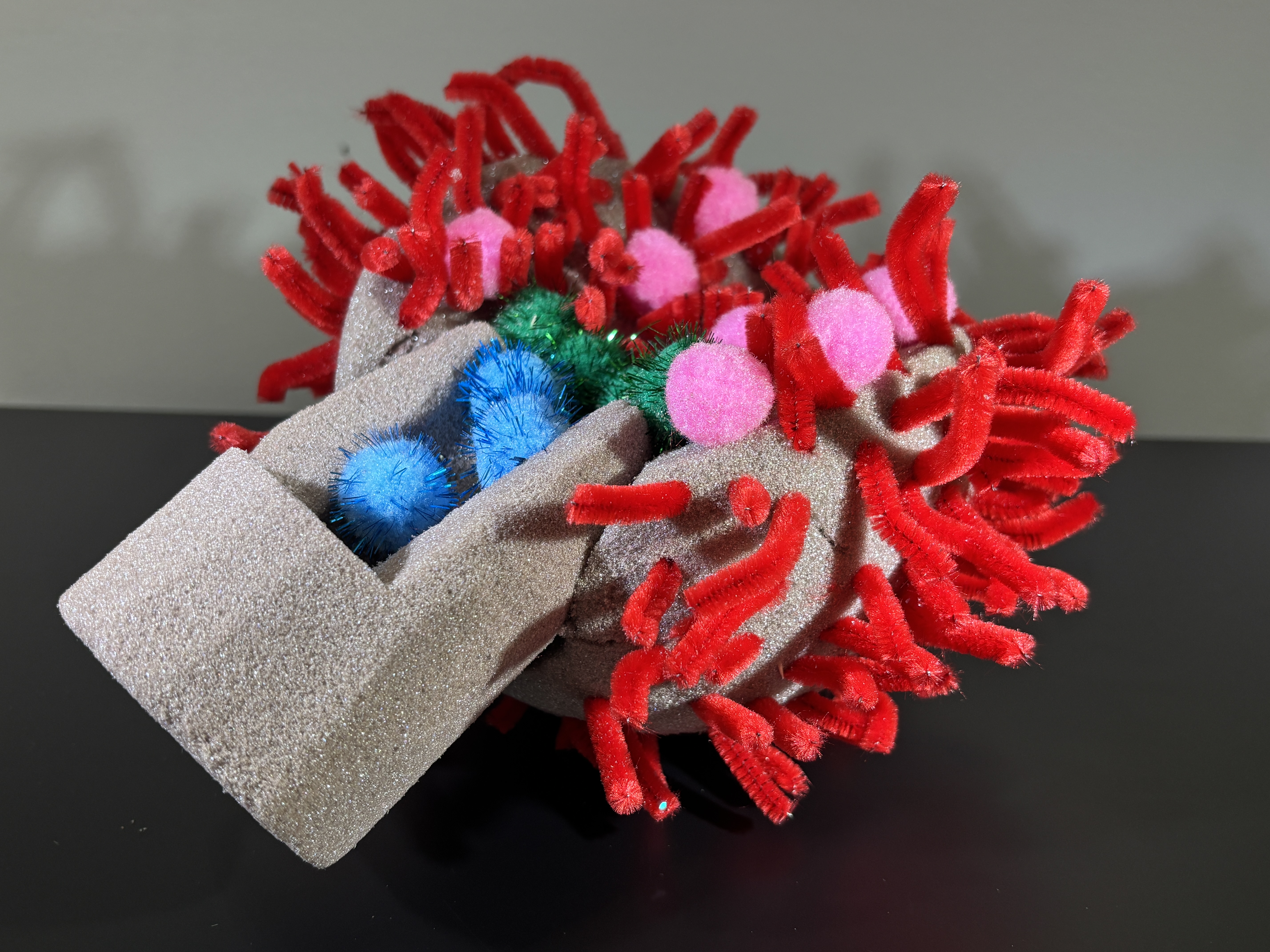
Root canal anatomy reveals a hidden world of remarkable complexity. Beneath the visible crown lies an intricate three-dimensional system of canals, curves, fins, accessory pathways, and apical deltas—each presenting unique clinical challenges. No two teeth are ever truly the same which keeps root canal treatment exciting and warrants its recognition as a distinct specialist discipline within Dentistry.
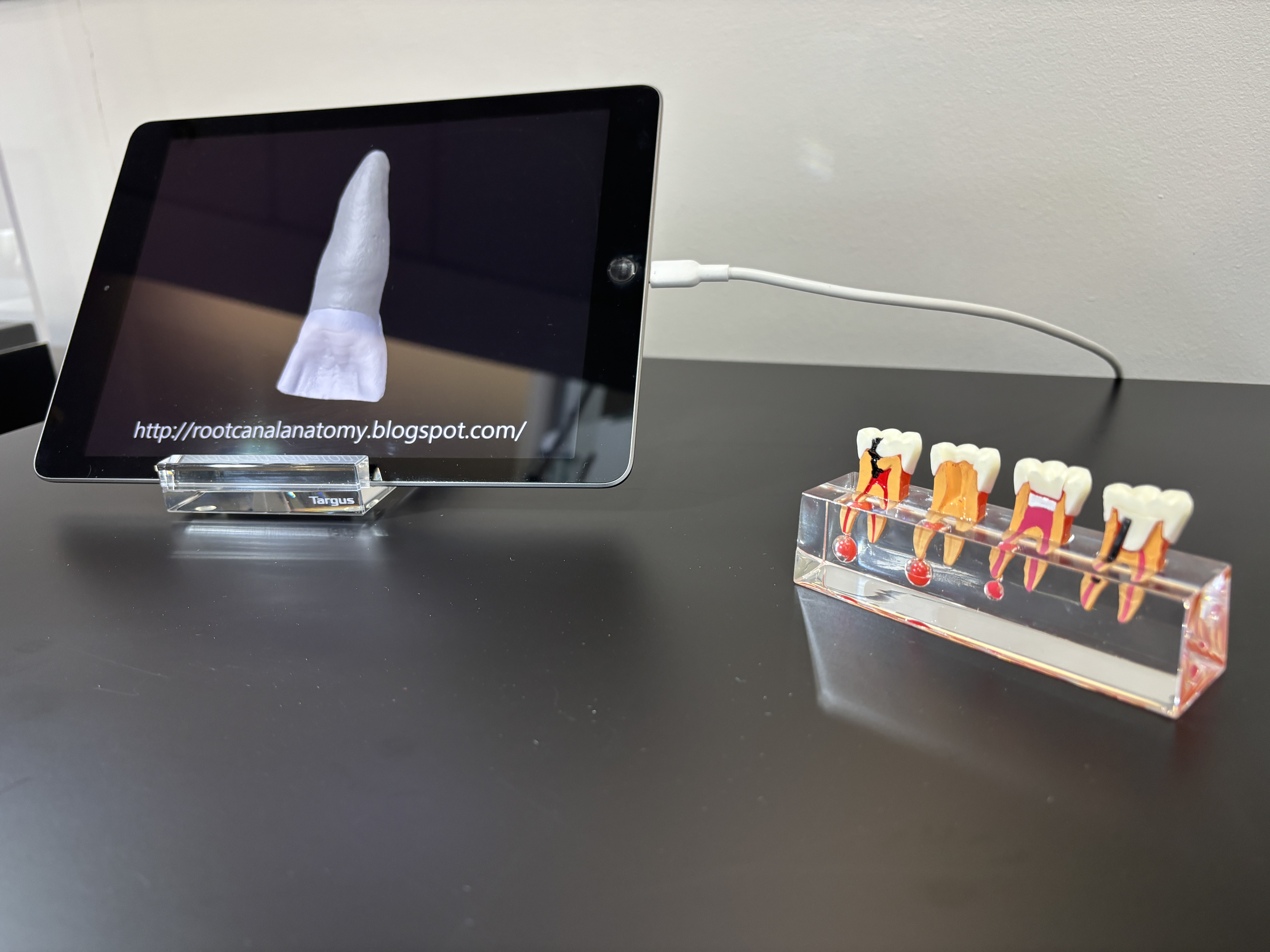
This exhibit brings that internal architecture into focus through a detailed anatomical model and dynamic visualizations (with permissions from Prof. Marco A. Versiani, allowing visitors to appreciate the spatial relationships and anatomical variations that define endodontic treatment.
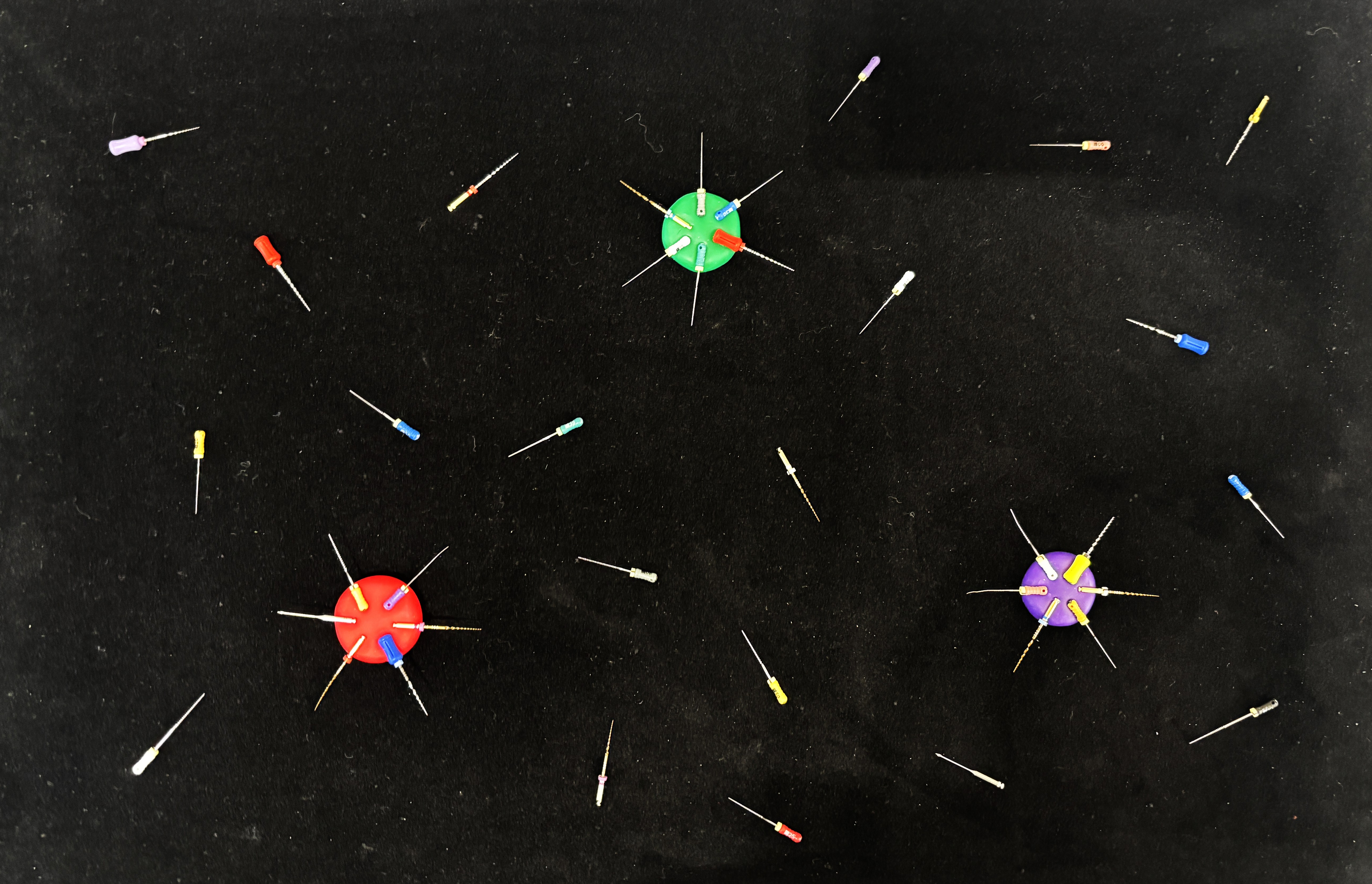
A thorough understanding of root canal morphology is fundamental to clinical success. Recognizing anatomical nuances enhances diagnostic accuracy, guides effective cleaning and shaping, and ultimately improves treatment outcomes. By exploring the architecture within, we deepen our respect for the precision, skill, and knowledge required for treatment modalities that preserve the natural dentition.

